96 Hour Chick Embryo Serial Section
This page on the embryology in chicken relates to the following: • Whole mount preparation 72 hours, with a detail view on the development of the head () • Cross sections 72 hours; eye and heart formation ( ) Stage 72 hours Whole-mount preparation 72 hours after fertilization Information: 72 Hours after fertilization, the rotation of the embryo to the left is arrived such behind the region of the heart and only the caudal part of the embryo must twist 90 degrees. The two flexures in the head region are almost completed. The fourth pharyngeal groove develops and the pharyngeal arches are thicker. Due to the cranial flexure, the pharyngeal region (= region of the trachea) is now located at the ventral side of the head. The fore and hind limbs at the level of the 16 th to the 20 th respectively the 27 th to the 32 th somite pairs are visible as small buds at an incubation time of about 3 days. Embryology of chicken 72 hours after fertilization: stained whole-mount preparation.  1 = Auditive (otic) vesicle, 2 = Myelencephalon, 3 = Metencephalon, 4 = Amnion, 5 = Mesencephalon, 6 = Optic vesicle + lens, 7 = Diencephalon, 8 = Epiphyse, 9 = Telencephalon, 10 = Branchial arches, 11 = Heart, 12 = Forelimb (wing) bud, 13 = Vitelline arteria/vein, 14 = Hindlimb (leg) bud, 15 = Tail Information: The development of the brain is shown at higher magnification.
Microscope Slide of Chick 96 hr. Serial cross section. Shows the beginning of most of the structures of the adult eye. Chick Embryo, 96 hour Microscope Slides. 24 hr chick Note that in the 24 hour chick, Hensen's node is located further caudally and the primitive streak is present only at the posterior end of the embryo. As Hensen's node retreats posteriorly, the organs which define the embryonic axis are formed anterior to it.
The further development of the five brain vesicles (telencephalon, diencephalon, metencephalon, mesencephalon and myencephalon) in to the different head structures is clearly visible. The eye vesicles differentiate as two lateral projections of the diencephalon and come in contact with the external layer (ectoderm) to form the optic cup (neurectoderm) and the lens (ectoderm). The dorsal projection of the dieencephalon is also visible and will differentiate in to the epiphysis (epi= at the upper side). The depression at the ventral side of the diencephalon develops in to the hypophysis ( hypo= under; not visible in this fig.). The auditory vesicles develop at the level of the myencephalon.
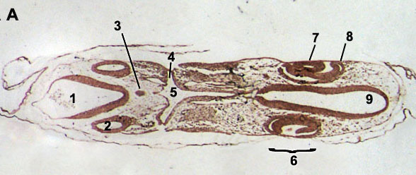
Embryology of chicken 72 hours after fertilization: detail of the head in a stained wholemount preparation 1 = Auditive (otic) vesicle, 2 = Myelencephalon, 3 = Metencephalon, 4 = Amnion, 5 = Mesencephalon, 6 = Optic bulb, 7 = Lens, 8 = Diencephalon, 9 = Epiphyse, 10 = Telencephalon, 11 = Olfactory groove Cross sections 72 hours A. Software ps2 pfs explorer download. Eye and heart formation: left wholemount and right cross section (dorsal part oriented to the left) 1 = Spine, 2 = Dorsal aorta, 3 = Foregut, 4 = Heart formation, 5 = Lens, 6 = Optic vesicle, 7 = Diencephalon, 8 = Heart region, 9 = head B. Heart formation: left wholemount preparation and right cross section (dorsal side oriented to the left) 1 = Spine, 2 = Dorsale aorta, 3 = Pleural cavity, 4 = Foregut with folding of the lung bud, 5 = Sinus venosus, 6 = Truncus arteriosus, 7 = Atrium, 8 = Regio cerebrale hemisphere of the telencephalon, 9 = Diencephalon, 10 = Olfactory groove, 11 = Heart region, 12 = Head region.